State-of-the-Art Technology

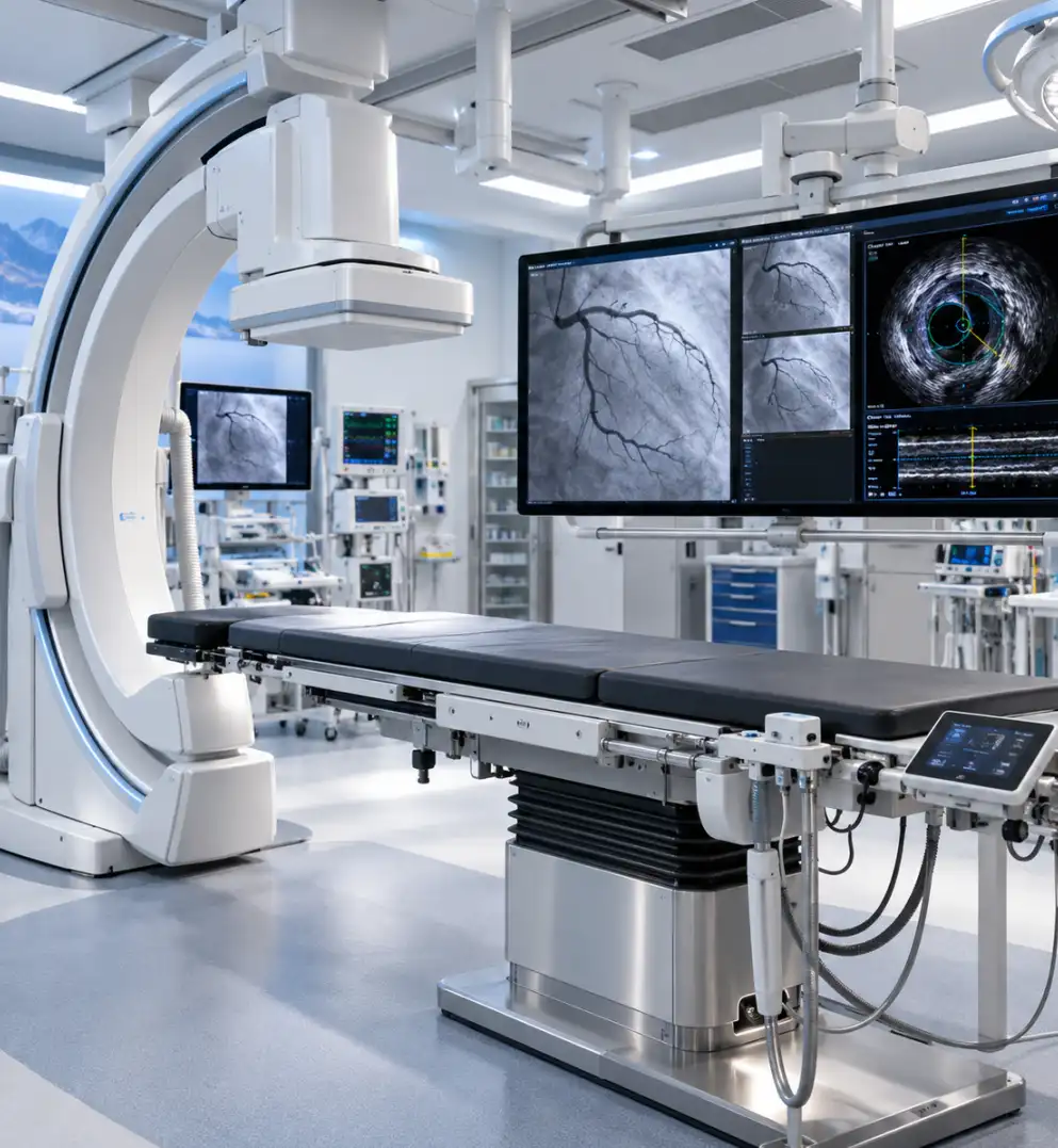
Cath Lab with IVUS

A cardiac catheterisation (cath) lab is a specialised procedure room with X-ray imaging equipment, a movable table, monitors and recording systems. Doctors pass thin catheters, usually through an artery in the wrist or groin, to the heart to diagnose and treat blocked arteries. Standard angiography injects contrast dye to outline the artery on X-ray. This shows the channel but not the vessel wall. IVUS (intravascular ultrasound) adds a miniature ultrasound probe at the catheter tip. Placed inside the artery, it produces cross-sectional images from within, showing the vessel's true diameter, the amount and type of plaque, calcium deposits and the layers of the wall. Doctors use this to choose stent size and length, confirm the stent has fully expanded against the wall, and assess unclear or complex blockages. Studies link IVUS-guided stenting to fewer repeat procedures and heart-related events than angiography alone, particularly in complex cases.
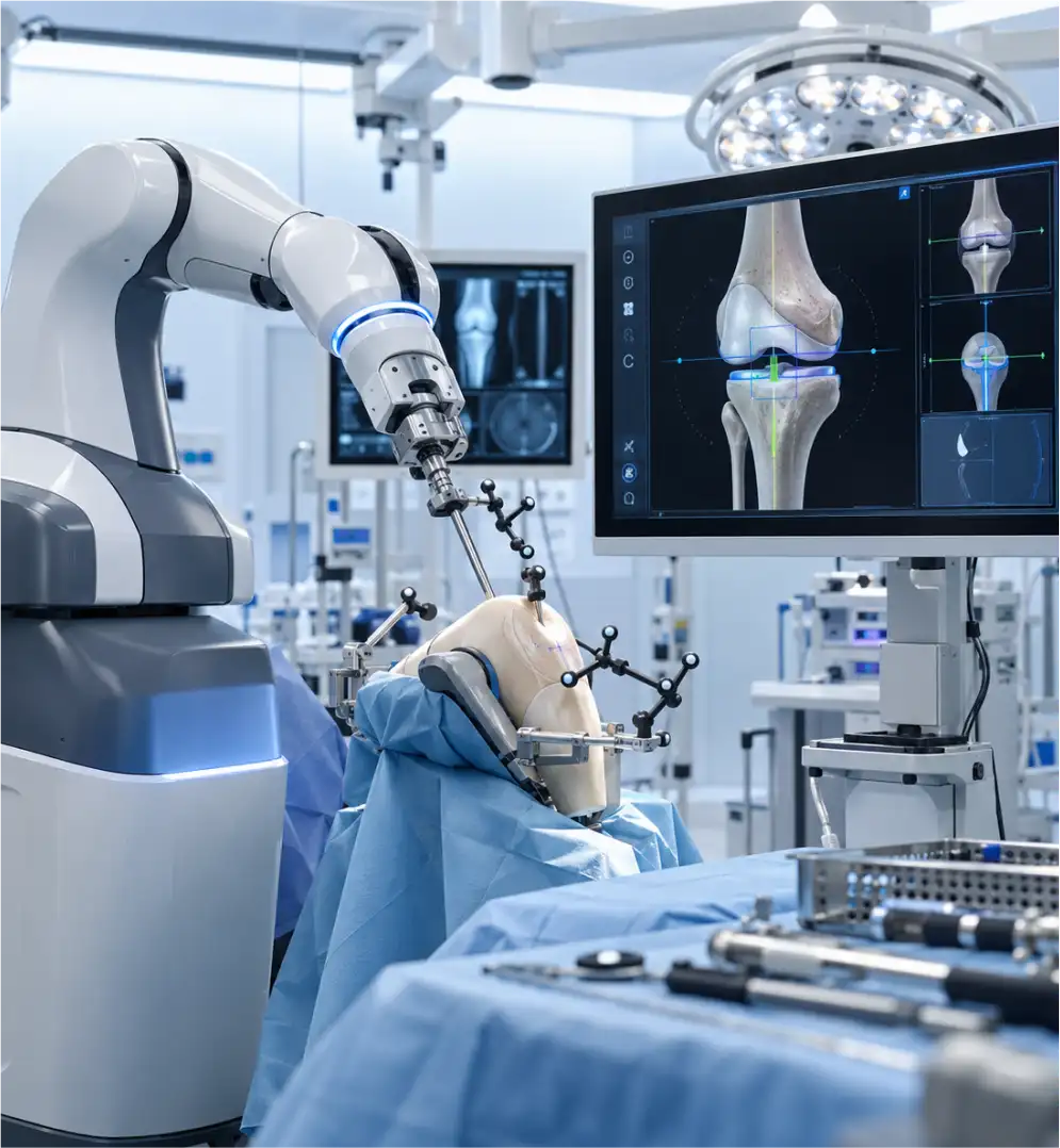
Robotic knee replacement

Knee replacement removes worn joint surfaces and replaces them with metal and plastic implants. Robotic-assisted systems are tools controlled by the surgeon, not autonomous machines. Most work in two stages. First, the knee is mapped, either from a pre-operative CT scan that builds a 3D model, or by recording bone landmarks during surgery. The surgeon uses this to plan implant size, position and alignment, and to balance the ligaments. During surgery, the system tracks the knee in real time and guides the cutting tool, often with boundaries that stop the saw or burr outside the planned area. Manufacturers describe accuracy within roughly a millimetre and a degree. Research shows more precise implant positioning, and some studies report less early pain and faster early recovery. Whether it improves long-term implant survival is still being studied. Drawbacks include higher cost and, initially, longer operating time.

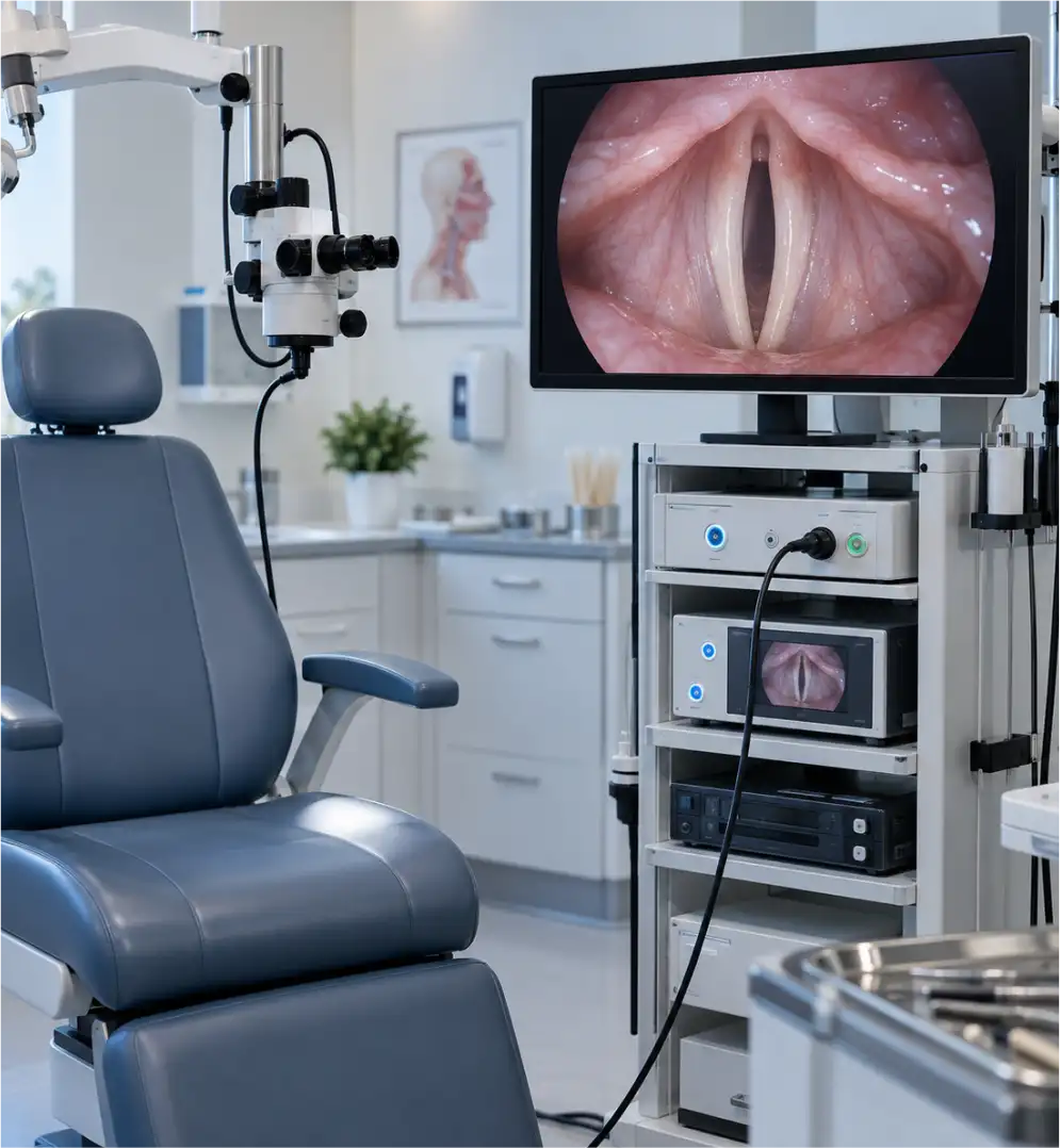
Stroboscopy

Stroboscopy, or videostroboscopy, is the standard clinical test for examining how the vocal folds vibrate. They open and close roughly 100 to 250 times per second, far too fast for the eye or ordinary video to follow. A rigid endoscope passed through the mouth, or a flexible one through the nose, carries a camera and a light that flashes in sync with the voice. The flashes are timed slightly out of step with the vibration rate, so each captures a different point in successive cycles. When played together, they create an apparent slow-motion view. Doctors assess the mucosal wave, symmetry, regularity and how fully the folds close. This helps identify nodules, polyps, cysts, scarring, paralysis and early tumours. It works best when vibration is fairly regular, and irregular voices may need high-speed imaging. The test takes a few minutes, is done awake, and may use a numbing spray.

Holmium laser

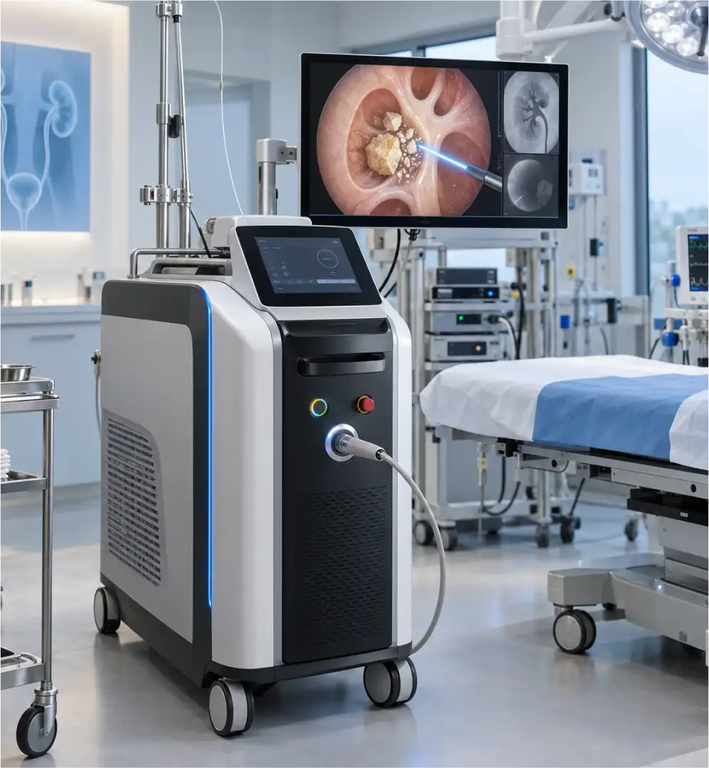
A holmium laser (Ho:YAG) is a pulsed laser that emits infrared light at 2,100 nanometres. Water strongly absorbs this wavelength, so the energy acts on a very small area, a fraction of a millimetre deep, which limits damage to nearby tissue. The light travels through a flexible optical fibre as thin as a hair, threaded through the working channel of an endoscope. In stone treatment (laser lithotripsy), pulses heat and break the stone into fragments or fine dust. Surgeons adjust pulse energy and frequency, using lower settings to produce dust and higher settings to break stones into pieces. The laser works on stones of any composition, including very hard ones. It is also used for prostate enlargement (HoLEP), urethral strictures and some bladder tumours. Safety measures include protective eyewear, continuous irrigation to cool the area, and careful fibre positioning. Rare risks include injury to the ureter or urethra.

Neuro microscope

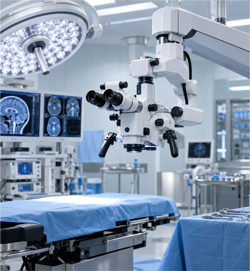
A neurosurgical operating microscope is a high-powered optical instrument that gives surgeons a magnified, well-lit, three-dimensional view of deep and delicate structures. Magnification typically ranges from about 4 to 40 times. Light travels along the line of sight, so narrow, deep corridors in the brain and spine stay illuminated without shadows. Motorised zoom and focus, and a counterbalanced arm that can be positioned and locked with a touch, let surgeons keep their hands on the instruments. Modern units add features such as fluorescence imaging, which highlights tumour tissue or shows blood flow in vessels, a second eyepiece for an assistant, integrated cameras, and links to navigation systems that overlay scan data on the view. Microsurgery with this technology has been standard in neurosurgery since the 1960s. It is used in brain tumour removal, aneurysm clipping, microdiscectomy and nerve repair.

Endoscopy suite

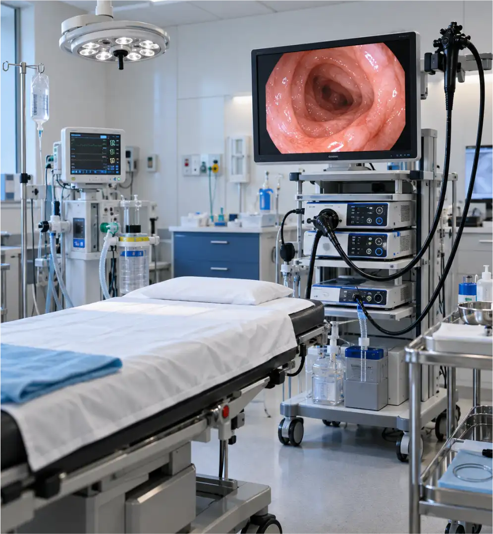
An endoscopy suite is a procedure area built around endoscopes, which are flexible tubes with a light source, a high-definition camera chip, and channels for air, water, suction and instruments. Images appear live on a monitor. Common procedures include gastroscopy (food pipe, stomach and first part of the small intestine), colonoscopy (large bowel), and ERCP and endoscopic ultrasound for the bile ducts, pancreas and deeper tissue layers. Imaging aids such as narrow-band imaging highlight surface patterns to help spot early abnormalities. Instruments passed through the scope can take biopsies, remove polyps, clip or band bleeding vessels, widen narrowings, place stents and extract bile duct stones. This lets diagnosis and treatment happen in one session. A suite also includes monitored sedation, carbon dioxide insufflation for comfort, and dedicated equipment for cleaning and disinfecting scopes between patients. Risks are low but include bleeding, perforation and sedation-related reactions.

Ureteroscope / RIRS / URSL

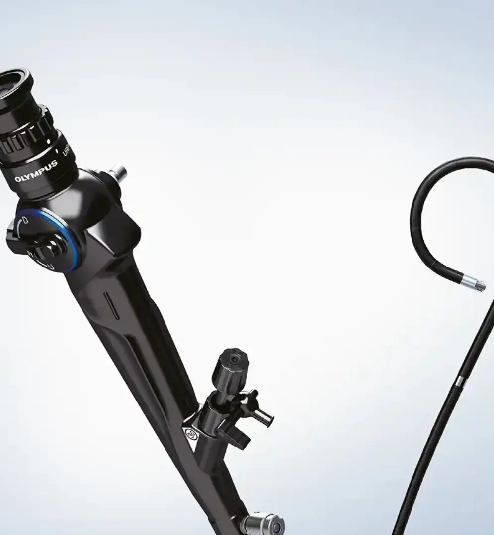
A ureteroscope is a thin telescope-like instrument, about 3 mm wide, with a camera at its tip and a narrow channel for tools. It is passed through the urethra and bladder into the ureter and, if needed, the kidney, so no incision is made. There are semi-rigid and flexible versions. Semi-rigid scopes suit stones in the ureter, a procedure called URSL (ureteroscopic lithotripsy). Flexible scopes bend sharply to reach inside the kidney's collecting system, which is called RIRS (retrograde intrarenal surgery). Stones are broken with a laser fibre, usually holmium, and fragments are removed with a tiny basket or allowed to pass. A thin sheath may protect the ureter, and a temporary stent is sometimes left in place afterwards. RIRS is generally used for kidney stones up to about 2 cm. The same scopes can examine strictures, bleeding sources and tumours in the urinary tract. Risks include infection, ureter injury and stent discomfort.

Mumbai's Strongest Healthcare Cluster.
Standing Tall for You.
Apex Group of Hospitals — Borivali, Kandivali and Mulund, united as one network for a healthier Maharashtra. Authentic, transparent healthcare, built around you and the people you love.